Cytology

Abnormal glandular cells
|
<<< Cervical cytology |
Abnormal glandular cells in cervical samples may be derived from the endocervix, endometrium, Fallopian tube and ovary. Benign glandular cells may show reactive changes due to inflammation, metaplasia and iatrogenic causes. Glandular cells can show atypia which may be borderline or dyskaryotic. Dyskaryosis in glandular cells is not graded.
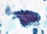
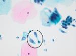
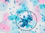
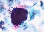
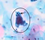
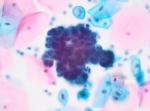
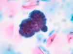
Glandular neoplasia of endocervical type (diagnostic code: 6 - ?Glandular neoplasia of endocervical type) is characterised by abnormal architecture of glandular cells as well as abnormal nuclei. Instead of the orderly honeycomb sheets and strips of epithelium with basally located nuclei in a normal sample, abnormal endocervical cells are seen in crowded sheets with overlapping nuclei and pseudostratified, crowded strips. The groups of cells often have "feathering" at the edges as the cells protrude from the edges of the group, and may form rosettes. The nuclei are enlarged and may appear to bulge out of the cytoplasm at the lateral borders of the cell, this is best seen when cells are present singly rather than in groups. The nuclei are usually hyperchromatic and may have irregular nuclear membranes. Samples from cases of endocervical glandular neoplasia often contain abundant glandular cells, including normal and abnormal forms.
Borderline changes in endocervical glandular cells may be diagnosed in some cases and this category is used in the same way as borderline changes in squamous cells: where the atypical features are not sufficient to diagnose dyskaryosis, but where dyskaryosis cannot be confidently excluded.
Where there is glandular neoplasia of endocervical type, always look for the presence of squamous dyskaryosis in addition as the two conditions can co-exist.
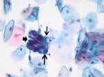
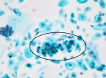
Other abnormal glandular cells (diagnostic code: 0 - Glandular neoplasia (non-cervical)) may be derived from the proximal tract. It is relatively rare to find these in cervical samples, but where they are identified, the most common origin is from an endometrial neoplasm. Abnormal endometrial cells show nuclear enlargement and may have nucleoli and irregular nuclear membranes. They usually have scanty cytoplasm, although they may contain vacuoles and sometimes have dense, blue-green squamoid cytoplasm. They may be seen containing phagocytosed neutrophils. Occasionally clusters of vacuolated ovarian carcinoma cells may be picked up in cervical samples.
Abnormal glandular cells may be present as hyperchromatic crowded groups which can be difficult to assess.
|
<<< Cervical cytology |